
GENERATIONS AHEAD
ALWAYS ADVANCING
MONTERIS CONTINUES TO LEAD with surgeons’ preferred technology platform. NeuroBlate® Fusion-S™ Software delivers visualization, control and efficiency needed to make critical decisions when performing brain tissue laser ablation, such as tumor, radiation necrosis and epilepsy procedures.
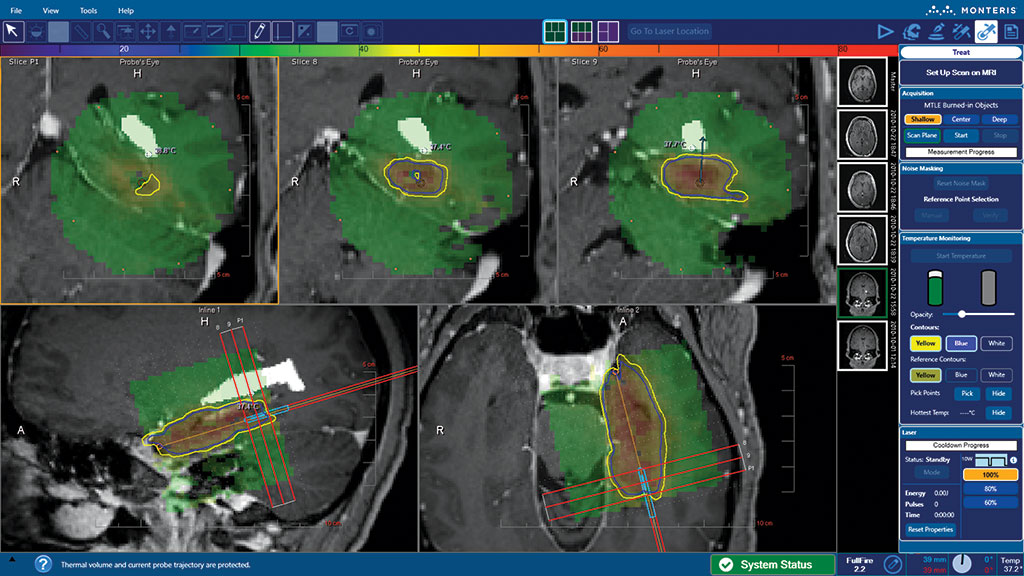
VISUALIZATION
NeuroBlate Fusion-S Software delivers superior visualization that allows surgeons to see more of the procedure to execute every procedure confidently.
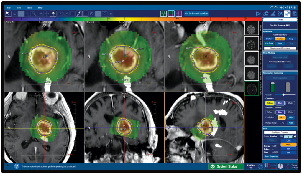
- 3-D anatomical views provide an advanced graphical interface to optimally define the ablation target and surrounding structures.
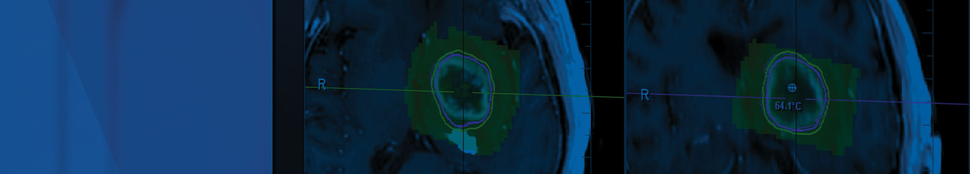
- Up to 2 thermal dose threshold (TDT) lines can be displayed simultaneously, allowing surgeons to tailor laser delivery that conforms to intended ablation boundaries.
CONTROL
Robust features provide the control and assurance you need to make critical decisions and manage the case from start to finish.
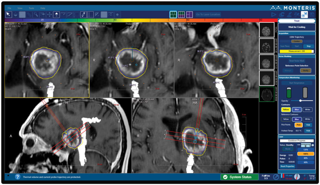
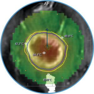
- Pick points can be assigned warning limits to ensure tissue temperatures remain within a safe range throughout the procedure
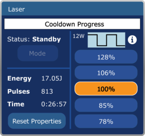
- Lower power settings enable precise control of energy delivery, which is especially beneficial while targeting small lesions.
EFFICIENCY
NeuroBlate Fusion-S Software includes features that allow for efficiency that is essential for today’s health systems.
- Shorter procedure times are possible with a reduction in reference scans before the ablation.
NeuroBlate Fusion-S Software. Generations Ahead.
Disclosures
Monteris provides technology for neurosurgeons, which allows them to ablate (destroy with heat), brain structures such as brain tumors, radiation necrosis, and epileptic foci. Monteris technology includes the NeuroBlate System, AtamA, and MiniBolt devices, which may be used together to apply the focused laser energy with little or no effect on surrounding healthy tissue. The NeuroBlate System provides clinicians a tool that offers near real-time control and MRI visualization of the therapy during laser ablation treatment.
All brain surgeries carry risk. Possible adverse events include, but are not limited to, hematoma, embolic events, edema, bleeding, cerebral spinal fluid (CSF) leakage, infection, unintended major tissue damage and permanent neurological deficits. Prior to using these devices, please review the Instructions for Use for a complete listing of indications, contraindications, warnings, precautions and potential adverse events.
Rx Only.